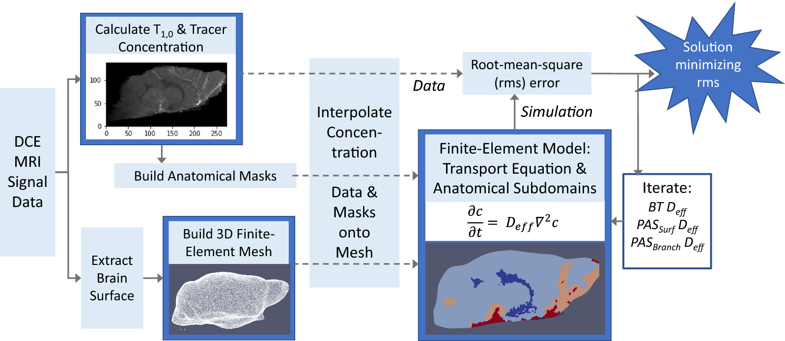
Fig. 2
From:Quantitative analysis of macroscopic solute transport in the murine brain

Schematic of brain transport analysis using finite-element modeling with DCE-MRI Data. DCE-MRI signal data is used to calculate contrast agent concentration and\(T_{1,0}\), a parameter used to identify different types of tissues. Contrast concentration and\(T_{1,0}\)are used to define anatomical masks for regions relevant to brain-wide transport, including the ventricular system, the macroscopic arterial vasculature, and its associated perivascular spaces. DCE-MRI data is also utilized to extract the surface of the brain. The anatomical masks are unique to each experimental subject. A 3D tetrahedral mesh is built from the brain surface. The concentration data and anatomical masks are interpolated onto the mesh. A finite-element model is developed utilizing the simplified mass transport equation (Eq.2) and subdomains defined by the anatomical masks (Fig.3). Unique effective diffusivities are applied to each subdomain. Simulations are performed for varying effective diffusivities to find the combination of effective diffusivities that minimizes the difference between the concentrations data and the simulation
